




Breast cancer survival comes down to one factor more than any other: when the cancer is found. According to the American Cancer Society, women diagnosed with localized breast cancer (cancer that has not spread outside the breast) have a five-year relative survival rate above 99 percent. For women whose cancer has spread to distant parts of the body before it is found, that rate drops to 33 percent. That gap is the entire argument for routine screening, and it is why a single mammogram appointment can be one of the most consequential 30 minutes a woman ever spends.
Screening matters because breast cancer is often silent in its earliest, most treatable stages. By the time a lump is large enough to feel during a self-exam, the cancer has usually been growing for some time. Mammograms find tumors years before they become physically noticeable, and that head start changes everything about treatment and outcomes.
This guide breaks down what every woman should know about breast cancer screening: when to start, what to expect, why early detection matters, and how to advocate for the right care. The information here is general. Your specific screening schedule should be set in conversation with your doctor based on your personal risk factors. For Atlanta-area women seeking guidance from a board-certified expert, Dr. Diane Alexander of Artisan Plastic Surgery, recently named lead medical expert in a Woman’s World feature on breast cancer screening, is part of our team of specialists committed to women’s health throughout every stage of life.
Why Early Detection Matters So Much
Breast cancer treated early is, in most ways that count, a different disease than breast cancer treated late. Survival rates and treatment options shift dramatically based on the stage at diagnosis.
The American Cancer Society reports the following five-year relative survival rates for women diagnosed with breast cancer between 2015 and 2021:
| SEER Stage | What It Means | 5-Year Relative Survival |
| Localized | Cancer is confined to the breast | Greater than 99% |
| Regional | Cancer has spread to nearby structures or lymph nodes | 87% |
| Distant | Cancer has spread to distant parts of the body, such as lungs or bones | 33% |
| All stages combined | Average across all diagnoses | 92% |
Source: American Cancer Society, Survival Rates for Breast Cancer (last revised January 2026). Based on SEER data from women diagnosed 2015-2021. Localized stage includes invasive cancer only and excludes ductal carcinoma in situ (DCIS).
The numbers tell a clear story. About 65 percent of breast cancers in the United States are diagnosed at the localized stage, when treatment is most effective and least invasive. The job of screening is to push that percentage higher by finding cancers before they have a chance to spread.
Beyond survival, early-stage cancers respond to less aggressive treatment. Lumpectomy, a breast-conserving surgery that removes the tumor and a small margin of tissue, is often an option for women whose cancer is caught early. That can mean avoiding a full mastectomy and the longer recovery that comes with it. According to the National Breast Cancer Foundation, women who receive regular screenings have a 26 percent lower breast cancer death rate than women who do not.
When to Start Breast Cancer Screening
Major medical organizations agree on the broad strokes of breast cancer screening, even if the specific schedules differ slightly. As of April 2024, the U.S. Preventive Services Task Force recommends that all women at average risk get screened with mammography every two years from age 40 to age 74.
The American Cancer Society’s guidelines recommend that women at average risk:
- Have the option to begin annual mammograms at age 40.
- Should begin annual mammograms by age 45 at the latest.
- Can transition to mammograms every two years starting at age 55, or continue annually based on personal preference.
- Should continue screening as long as they are in good health and have a life expectancy of 10 or more years.
Women at higher than average risk should start earlier. Higher risk includes:
- Family history. A first-degree relative (mother, sister, daughter) diagnosed with breast or ovarian cancer, especially before age 50.
- Genetic mutations. Known BRCA1, BRCA2, or other inherited mutations that increase breast cancer risk.
- Personal history. A previous breast cancer diagnosis, atypical hyperplasia, or lobular carcinoma in situ (LCIS).
- Radiation exposure. Prior chest radiation, often for treatment of childhood or young-adult cancers like Hodgkin lymphoma.
- Dense breast tissue. Categories C and D on a mammogram report, which both increase cancer risk and make tumors harder to spot.
Women in any of these categories should talk with their doctor about beginning screening as early as age 30, often combined with breast MRI for additional sensitivity. The standard age guidelines exist for women without these risk factors. They are a floor, not a ceiling.
What to Expect at a Mammogram

A screening mammogram takes about 20 to 30 minutes from check-in to walking out the door. The actual imaging takes only a few minutes.
During the appointment:
- Check in and change. You will change into a gown that opens in the front. Avoid wearing deodorant, lotion, or powder on screening day, since these can show up on the images.
- Imaging. A radiologic technologist positions each breast on a flat plate. A second plate compresses the breast briefly while images are captured. Most facilities take two views of each breast.
- Discomfort, not pain. Compression is brief, usually 10 to 15 seconds per view. Some women find it uncomfortable, especially if their breasts are tender, but it should not be sharply painful.
- Results. A radiologist reviews the images, usually within a few business days. Most results come back normal. If something needs a closer look, you will be called back for diagnostic imaging, which is common and often turns out to be benign.
Booking is easier than many women expect. Most imaging centers accept self-referrals, and screening mammograms are covered with no out-of-pocket cost under most insurance plans, including Medicare.
2D vs. 3D Mammography: What’s the Difference?
Standard 2D mammography produces flat images of the breast, similar to a single photograph. 3D mammography, also called digital breast tomosynthesis, takes multiple low-dose images at different angles and combines them into a layered view. A radiologist can scroll through the breast tissue layer by layer, almost like turning the pages of a book.
The clinical benefit shows up in the numbers. A study published in Radiology comparing 3D and 2D mammography across more than 77,000 patients found that 3D digital breast tomosynthesis improved overall cancer detection by 28.6 percent compared with 2D mammography, while reducing recall rates (the rate of false alarms) by 16.1 percent. For women with dense breast tissue, where standard mammograms can miss tumors hiding behind glandular tissue, the difference is even more significant.
Most major imaging centers in Atlanta now offer 3D mammography as a standard option. If your insurance covers it, it is worth asking for.
What Dense Breast Tissue Means for Screening
According to the FDA, approximately half of women over 40 in the U.S. have dense breast tissue, meaning their breasts contain more glandular and fibrous tissue and less fatty tissue. Density is a normal anatomical feature, not a problem in itself, but it has two consequences for screening:
- Higher risk. Women with dense breasts have a higher risk of developing breast cancer than women with mostly fatty tissue. Research suggests women with extremely dense breasts have about four times the risk compared with women whose breasts are mostly fatty.
- Harder to read. On a 2D mammogram, dense tissue appears white. So do tumors. The overlap can mask cancers, creating a ‘frosted glass’ effect that obscures small abnormalities. About 40 percent of breast cancers can go undetected on standard mammography in extremely dense breasts.
As of September 10, 2024, federal law (under the FDA’s updated Mammography Quality Standards Act) requires that mammogram reports include information about breast density. Every woman who gets screened in the United States now receives this information. If your report says you have dense breasts (categories C or D), ask your doctor about supplemental imaging. Options include:
- 3D mammography (digital breast tomosynthesis)
- Breast ultrasound
- Breast MRI, especially for women at high risk
- Molecular breast imaging, available at some specialty centers
The right combination depends on your overall risk profile. A primary care doctor or OB-GYN can help calculate that risk and refer you to the right imaging.
How to Advocate for Yourself
In her recent Woman’s World feature on breast cancer screening, actress Regina Hall delivered a clear message to women: advocate for yourselves. The medical experts in the article, including Dr. Diane Alexander, agreed.
Self-advocacy in this context means a few specific things:
- Know your family history. Sit down with your relatives and document who in your family has had breast, ovarian, prostate, or pancreatic cancer, and at what age. This information shapes your screening plan.
- Ask about your breast density. Your mammogram report will now state your density category. If it is unclear, ask your doctor what it means for you.
- Push for the right imaging. If you have dense breasts or other risk factors, supplemental imaging is not a luxury. It is appropriate care, and a growing number of insurance plans now cover it.
- Trust your body. If something feels different, a new lump, skin changes, nipple discharge, persistent pain, do not wait for your next scheduled mammogram. Call your doctor.
- Make screening routine. Put it on the calendar at the same time every year, the way you would an annual physical. Removing the decision removes the excuse.
Where a Plastic Surgeon Fits In
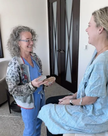
Plastic surgeons are part of breast cancer care from the very beginning, not just at the end. Reconstruction options shape the conversations a woman has with her oncologic surgeon, and that conversation should ideally happen before treatment decisions are finalized.
Women diagnosed with breast cancer have several reconstruction paths to consider:
- Immediate reconstruction. Performed during the same surgery as the mastectomy or lumpectomy.
- Delayed reconstruction. Performed weeks, months, or even years after cancer treatment is complete.
- Implant-based reconstruction. Uses saline or silicone implants to rebuild breast volume.
- Autologous reconstruction. Uses the woman’s own tissue, often from the abdomen, back, or thighs, to rebuild the breast.
Knowing what reconstruction can look like before cancer surgery happens helps a woman make more informed choices about her treatment. Dr. Alexander and the team at Artisan Plastic Surgery work alongside oncologic surgeons across Atlanta to support patients through every stage of breast cancer treatment and recovery.
Frequently Asked Questions
How often should I get a mammogram?
The U.S. Preventive Services Task Force recommends biennial (every two years) screening from age 40 to 74 for women at average risk. The American Cancer Society recommends annual screening starting at age 40 to 45. Women with a family history of breast cancer, a known genetic mutation, or other risk factors may need to start earlier and screen more often. Talk with your doctor about the right schedule for you.
Are mammograms safe?
Yes. The radiation dose from a screening mammogram is low, roughly equivalent to the natural background radiation a person absorbs over about seven weeks. The benefits of finding cancer early far outweigh the small risk from imaging.
What if I have dense breasts?
Dense breast tissue makes mammograms harder to read and increases breast cancer risk. As of September 2024, your mammogram report is required by federal law to indicate whether your breasts are dense or not dense. If your report indicates dense breasts (category C or D), ask your doctor about supplemental imaging such as 3D mammography, ultrasound, or MRI.
Do self-exams replace mammograms?
No. Self-exams help women learn what is normal for their bodies, which makes it easier to notice changes, but they cannot find tumors as small as a mammogram can. Both are part of good breast health, and neither replaces the other.
What does Dr. Alexander recommend for women in Atlanta?
Dr. Alexander’s guidance, consistent with her published commentary in Woman’s World, is straightforward: make screening routine, know your density and family history, and do not wait for symptoms to act. Women with concerns about breast health, reconstruction, or cosmetic procedures can schedule a consultation with her at Artisan Plastic Surgery.
The Bottom Line
Breast cancer screening is the single most effective tool for catching breast cancer early, when treatment is most successful. The data is clear and consistent: women who screen regularly survive at higher rates than women who do not. Stage at diagnosis matters more than almost any other factor, and screening is what determines stage.
If you have not had a mammogram in the past year, the next step is simple. Call your doctor or a local imaging center and book one. If you have questions about your risk, your density, or your options for reconstruction or breast surgery, it helps to consult with an Atlanta plastic surgeon who will take the time to answer them.
At Artisan Plastic Surgery, Dr. Diane Alexander and our team work with women across Atlanta on every stage of breast health, from reconstruction after cancer treatment to cosmetic breast procedures. If a cosmetic procedure is part of your journey, our guide to what a breast augmentation actually involves walks through the details. Schedule a consultation to learn more about our breast services and how we can support you.
Learn More
Read the full Woman’s World feature with Dr. Alexander: Regina Hall Urges Women to Get Breast Cancer Screenings: ‘We Have to Advocate for Ourselves’.
See Dr. Alexander’s verified author profile and additional contributions on the publication’s site: Diane Alexander, MD, FACS at Woman’s World.
Sources
American Cancer Society. Survival Rates for Breast Cancer (last revised January 13, 2026): cancer.org/cancer/types/breast-cancer
American Cancer Society. American Cancer Society Recommendations for the Early Detection of Breast Cancer: cancer.org/cancer/types/breast-cancer/screening-tests-and-early-detection
U.S. Preventive Services Task Force. Final Recommendation Statement: Breast Cancer Screening (April 2024): uspreventiveservicestaskforce.org/uspstf/recommendation/breast-cancer-screening
National Cancer Institute SEER Program. Cancer Stat Facts: Female Breast Cancer: seer.cancer.gov/statfacts/html/breast.html
U.S. Food and Drug Administration. Mammography Quality Standards Act Final Rule (effective September 10, 2024): fda.gov/radiation-emitting-products/mammography-quality-standards-act
Friedewald SM, et al. Clinical performance metrics of 3D digital breast tomosynthesis compared with 2D digital mammography. Radiology, 2014: pubmed.ncbi.nlm.nih.gov/24918774
National Breast Cancer Foundation. Breast Cancer Facts & Statistics 2026: nationalbreastcancer.org/breast-cancer-facts
Susan G. Komen Foundation. Understanding Breast Cancer Survival Rates: komen.org/breast-cancer/facts-statistics/breast-cancer-statistics/survival-rates

